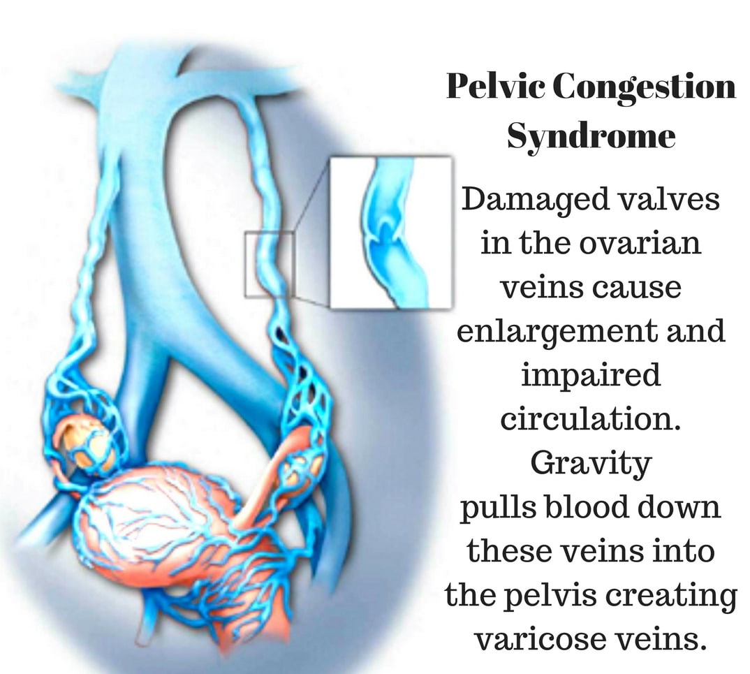
Pelvic Venous Congestion Syndrome is a chronic disease manifested by prolonged pain in the lower part of the trunk. This disease, which is also translated as Pelvic Obstruction Syndrome, is known as uterine varicose.
It is caused by the occlusion of the blood vessels in the pelvis and the accumulation of excess blood in this area. The pelvis is a structure located in the lower part of the body.
It is intertwined with different organs and systems. It is associated with the reproductive, urinary and digestive systems. And it supports the lower part of our body.
It carries almost half of a person’s body weight. There is no definite information about its incidence in the population. However, 1 out of 10 patients presenting with long-term abdominal pain has pelvic obstruction.
The most basic cause of Pelvic Obstruction Syndrome is the presence of obstruction and dilatation in the pelvic veins (retroaortic right and left renal vein).
“Pelvic Congestion Syndrome”, known among the people as “expansion of the uterine vessels”, can occur for many different reasons. The majority of people with this syndrome first complain of a blunt pain that radiates to the groin or groin and causes great discomfort.
As a result of the blockages in the pelvic region, as I described above, some symptoms will occur in people.
A number of complications can be seen in patients who are not intervened or treated in a timely manner.
The diagnosis of pelvic congestion syndrome can be made easily with advanced imaging methods such as computed tomography, ultrasound, and transvaginal ultrasonography in addition to the examination.
The backflow of blood in the ovaries and pelvic veins due to insufficient valves is the main problem in the occurrence of pelvic congestion syndrome. However, the emergence of this syndrome may also be due to obstructive anatomical conditions and diseases.
Another factor that causes “Pelvic Congestion Syndrome” is “multiple birth“. Due to the vasodilating effects of estrogen and progesterone hormones, “expansions” or “varices” occur in the abdominal and leg veins both due to hormonal reasons and due to the pressure of the baby in the mother’s womb.
This situation occurs within a certain period of time. And it usually develops due to the deterioration of the “venous valve mechanism“, that is, the “valves” in the vein.
However, the weight gained during pregnancy and the anatomical changes in the pelvic structures due to pregnancy directly affect the venous blood flow in the pelvic region.
The blood flow to healthy veins slows down due to the accumulation of blood in the pelvic and ovarian veins. And it even comes to a standstill.
From this perspective, “pelvic pain” is usually caused by blood clotting in the vein (thrombosis) and the pressure exerted by the enlarged veins on nearby nerves.
The compression of the right common iliac artery to the left common iliac vein.
It is the condition where the left renal vein is located behind the aorta and the left ovarian vein is exposed to pressure.
It is a condition in which the left ovarian and left renal vein is compressed by the superior mesenteric artery.
Diagnosis of Pelvic Congestion Syndrome is still quite difficult, even if it is in a shorter and easier position compared to past times.
However, the diagnosis of the syndrome can be made at an earlier period in patients with highly significant varicose vasodilation in the vulva or vagina.
In addition to this condition, more than half of patients with pelvic congestion syndrome have ovarian cysts.
However, although the reason for this connection between pelvic congestion syndrome and the formation of ovarian cysts cannot be fully explained, it is thought that the connection arises due to excessive stimulation of the estrogen hormone.
This examination can be done both “transabdominal”, that is, “through the abdomen” and “transvaginal”, that is, “through the vagina”. The contribution of “doppler ultrasonographic examination” to the detailed examination of the venous blood flow in the pelvic region is quite large.
In order to perform venographic examination, imaging devices used in the angio department are needed. Usually, the right inguinal vein is used to perform this operation.
With the help of a wire and a catheter (plastic tube) placed in the inguinal vein, the left and right ovarian veins can be seen separately. In this way, the diameters of the vessels and the direction of blood flow in them are determined.
It can be called a drug in early stage patients. Some drugs that will prevent vasodilation and restore hormone balance can reduce the rate of progression of the disease. And sometimes it can stop progress. However, various pain relievers are used to relieve pelvic pain.
However, the most important technique in the definitive treatment of the disease is embolization of problematic pelvic vessels with a catheter.
In these procedures performed from the inguinal or neck vein, enlarged, structurally impaired vessels are detected in the pelvic region. These problematic veins are closed with special equipment.
The technical success of the pelvic venous embolization procedure is 99%, and the recurrence rate is below 10%. Transactions can be made in any season of the year. Patients can be discharged on the same day and continue their work and social life the next day.
The timing of the procedure has nothing to do with the menstrual period. There is no change in fertility and menstrual pattern after the procedure.
What is Varicocele? Varicocele is the varicose veins that drain the blood in the testicles,…
What is Hemorrhoids ? It is a disease caused by the loosening of the veins…
What Is Monkeypox Virus? Symptoms and Ways of Transmission! The monkeypox virus, which has been…
What is Myoma ? Myoma, is a benign tumor arising from the uterine muscles. It…
What is Back Lift? Back stretching, excessive weight gain and aging may cause you to…
What is a Gastric Balloon? Gastric balloon (gastric balloon) is one of the obesity treatments…