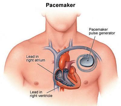
Heart Battery (Pacemaker) are state-of-the-art electronic devices that can provide electrical stimulation to the heart when necessary. It consists of battery, generator and cables. The metal box containing the electronic circuits that provide energy to the battery and manage the operation of the battery is called a generator.
The electrical communication between the heart and the generator is provided by cables called “lead”. The Heart Battery (Pacemaker) detects the electrical signals coming from the heart through these cables and, when necessary, stimulates the heart through these cables.
A Heart Battery (Pacemaker) is inserted because the heart’s stimulation center, namely the sinus node, cannot generate enough stimulation, the heart rate slows down, and the patient can continue to live accordingly.
Heart Battery (Pacemaker) is generally placed in people who have heart rhythm disorder and conduction disorder in the heart. People with these problems may have a much slower or faster pulse than normal, and the person’s heart may also have trouble pumping enough blood. The pacemaker is the most effective tool used to solve these problems and significantly improves a person’s quality of life.
The Heart Battery consists of a generator that produces electrical impulses and an electrode that transmits electrical impulses. Generators carry batteries containing lithium. These batteries are placed inside the right or left chest wall or in the abdomen.
Temporary Pacemakers are devices that are inserted to be removed when the patient’s heartbeat returns to normal.
It enters from the neck, lower part of the collarbone, arm or groin area.
And this Pacemaker, which consists of a cable reaching the heart and a small box located on the outside of the body, can be easily removed when necessary.
Generally, permanent pacemakers placed under the collarbone and just under the skin are preferred in the treatment of more chronic diseases such as atrioventricular block, tachycardia, bradycardia, hypertrophic cardiomyopathy and congestive heart failure.
Permanent batteries, which create the necessary stimulus for the heart to contract and relax, are classified in 3 different ways according to the stimulated region of the heart:
It is a type of Heart Battery that consists of a single cable and is used to stimulate only the right atrium or right ventricle of the heart.
The batteries used to stimulate both the right ventricle and the right atrium of the heart are called two-chamber Pacemakers.
One of the two separate leads is placed in the right ventricle. The other is placed in the right atrium. And when necessary, contraction is provided in both the ventricle and the atrium.
This type of battery, also called a biventricular Heart Battery, is known as the Cardiac Reconstruction Therapy Battery (CRT-P) and has an important form of warning needed for the treatment of heart failure.
Thanks to three different cables placed in the right atrium, right ventricle and left ventricle regions of the heart, it is ensured that even the heart muscle, which has lost its strength, contracts correctly and that each chamber of the heart works synchronously.
Pacemaker application is performed in sterile operating room conditions or in catheter laboratories. One day before the procedure, the patient’s intervention areas such as chest, armpit and groin are shaved. And each area is thoroughly cleaned with foam sponges. Then, the patient is taken to a sterile environment for the procedure and the area to be treated is cleaned by brushing with disinfectant solutions for 10 minutes just before the procedure.
The patient, who is brought to the appropriate position for the intervention, is covered with a dry sterile cloth. And the process begins in the operating room environment. Although regional anesthesia is generally preferred during pacemaker placement, heavy sedation or general anesthesia may be required in some cases.
Veins in the patient’s neck, armpit, arm or just below the collarbone are preferred for intervention. The pacemaker vein is entered through the catheter and the electrode part of the pacemaker is visualized with a fluoroscopy device and advanced to the heart.
The box-shaped part of the device, called the generator, is most likely located inside the right or left chest muscle. Then, the incisions made in the vein and muscle tissue are closed and the process is terminated. And if necessary, the patient is taken to the recovery unit.
General anesthesia is not required in most cases when a pacemaker is inserted. The area where the pacemaker will be inserted is anesthetized with local anesthesia.
Assistance is obtained from an x-ray device that constantly monitors the surgical site from the inside and monitors the condition of the electrodes in the body. During the procedure, a large vein connected to the heart is selected.
An electrode is placed in the heart through this vein. Which vein to choose may vary depending on the condition of the person. The most commonly used veins in this procedure are the femoral vein in the groin, the subclavian vein in the shoulder region, and the jugular vein in the neck.
Implantation of permanent pacemakers and ICD devices is usually done in a similar way. The surgery is performed in a fully sterilized operating room or clinic.
Local anesthesia is used, such as a temporary pacemaker. The presence of scopic imaging devices is of great importance during the preparation and operation of the pacemaker placement procedure. With these imaging methods, the area where the pacemaker will be inserted is carefully monitored and a point shot is made.
A small incision is then made just below the person’s collarbone. The size of this cut is about 3 cm. The “generator”, which is part of the pacemaker device, is inserted through this incision. Permanent pacemakers may have more electrodes than temporary pacemakers.
These electrodes are placed in the chamber(s) of the heart through a vein running just below the collarbone. Electrodes are placed in the chambers of the heart. And then the connection between the generator and the electrode is established. The incision is closed and the operation is terminated.
Pacemaker implantation is a smaller procedure compared to other surgical operations. Therefore, the occurrence of unwanted complications in the person is very unlikely.
Complications that may occur after surgery are generally low-risk and do not reach a level that threatens the person’s life.
Although very rarely, there may be cases such as rupture of the membrane layer in the lung during the incision during the surgery, accidental entry into the artery instead of the vein.
Although it is not very common, sometimes the area where the operation was performed may become infected after the patient is discharged. To eliminate this possibility, antibiotic drugs are usually given to the patient after surgery.
Sometimes, the generator and electrodes connected to the pacemaker can come out of the skin. In this case, the operation area is opened again. And the pacemaker is repaired.
What is Varicocele? Varicocele is the varicose veins that drain the blood in the testicles,…
What is Hemorrhoids ? It is a disease caused by the loosening of the veins…
What Is Monkeypox Virus? Symptoms and Ways of Transmission! The monkeypox virus, which has been…
What is Pelvic Venous Congestion Syndrome? (Failures Observed in Ovarian/Testicular Veins) What is Pelvic Venous…
What is Myoma ? Myoma, is a benign tumor arising from the uterine muscles. It…
What is Back Lift? Back stretching, excessive weight gain and aging may cause you to…